
Which tool is best for school screening?
Traditionally images of the cornea were taken by shining rings of light (Palcido Discs) onto the tear film on the surface of the eye. Then videoing how the reflections of these rings of light change when reflected off the tear film. This was further facilitated by using one or two cameras to take additional images of the eye.
In 2017 a new method to image the cornea using a laser was released onto the medical market. This device called the Anterion uses swept source optical coherence tomography (SS-OCT). We have used both devices in our clinic. Prior to using the swept source optical coherence tomography, we used a G4 device. This had the Placido disc images and dual rotating cameras.
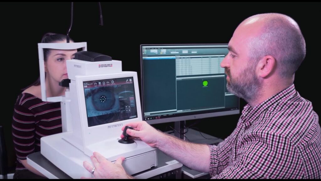
We had to take 3 images of each eye, the operator then had to review the quality of each image and select the best for analysis. Relative to the SS-OCT, this required a lot of time and storage for all the data generated. With our new Anterion I can capture an image within 20 seconds and the device automatically detects the image quality. If poor quality, it rejects the image, and another attempt needs to be taken. It is not reliant of the reflection off the tear film – a great source of poor-quality images with our previous device. All our Tech’s LOVE the new SS-OCT because of its speed of image taking, the fact that we only need one scan per eye and that it immediately performs image quality assessment and auto-rejects poor quality images.

Compared to Placido disc-based devices like the Pentacam, SS-OCT systems, such as the ANTERION by Heidelberg Engineering, offer a simpler, more efficient, and comprehensive approach to imaging the cornea.
Swept-Source OCT: A Simpler and More Efficient Process
SS-OCT uses a high-speed, tunable laser that “sweeps” across different wavelengths to capture detailed images of the eye’s structures. One of the primary advantages of this method is its ability to capture not just the surface of the cornea but also the deeper layers, providing a full thickness map of the cornea. Devices like the ANTERION integrate SS-OCT into a streamlined workflow, making the process both simple and efficient for clinicians and patients alike.
In terms of simplicity, SS-OCT offers several key advantages:
- Fast Acquisition
- Non-Invasive and Contact-Free
- Comprehensive Corneal and Anterior Segment Imaging
- Automated, User-Friendly Interface
- Excellent SCORE value that can safely rule out if someone has keratoconus.
1. Fast Acquisition:
SS-OCT systems capture thousands of A-scans per second, resulting in rapid acquisition of 3D images. This reduces the time the patient needs to be in front of the machine, which is particularly useful in busy clinical settings. With the ANTERION, the scanning process takes only a few seconds, minimizing patient discomfort and movement that could affect image quality.
The ANTERION and other SS-OCT systems are designed with automation in mind.
2. Non-Invasive and Contact-Free:
Like the Pentacam, SS-OCT is non-contact, which means there’s no need for corneal dyes or direct contact with the eye, reducing the risk of discomfort and infection. However, SS-OCT offers an additional advantage in that it can image through ocular opacities better than the Pentacam, leading to clearer results in patients with early-stage corneal haze or scarring.
3. Comprehensive Corneal and Anterior Segment Imaging:
SS-OCT provides more than just a corneal topography map. It captures the entire anterior segment, including the cornea, iris, and anterior chamber, in a single scan. This gives the clinician more data to work with, especially when considering the overall eye structure in keratoconus patients. Placido-based devices like the Pentacam, in contrast, are limited to corneal topography, and while they are excellent at measuring corneal curvature, they do not provide the same level of depth or detail regarding the internal corneal layers.
4. Automated, User-Friendly Interface:
- After positioning the patient, the system automatically captures and processes the images, requiring minimal input from the operator.
- This contrasts with the Pentacam, which requires more precise alignment to ensure accurate Placido disc imaging
- The automated nature of SS-OCT reduces the learning curve for technicians and lowers the chances of user error, making it simpler to implement in various clinical settings.
5. SCORE value:
This has high selectivity in detecting patients without keratoconus. After screening a patient there is a single SCORE value that if greater than Zero, will indicate that there is a good probability that a child may have keratoconus. Pentacam also has a well-researched and validated scoring system that can be used to detect patients without the condition. It however requires a visual acuity test to be added to the scoring system. With screening in schools, measuring a child’s vision will add on a significant total time spent testing each and every child.
Pentacam: Still a Reliable Tool but with Limitations
The Pentacam, based on Scheimpflug imaging and Placido disc technology, has long been a trusted device for corneal tomography. It is particularly effective in measuring corneal curvature and elevation and has been used extensively in diagnosing and monitoring keratoconus. However, its reliance on reflected light from the corneal surface limits its ability to capture detailed images of deeper corneal structures.
Another limitation of the Pentacam is its sensitivity to eye movement and tear film irregularities. The Placido disc technique depends on the clarity and smoothness of the corneal surface. Any irregularities, such as dry spots or minor eye movements during the scan, can distort the images. In contrast, SS-OCT’s high-speed scanning and ability to penetrate deeper structures make it less susceptible to such issues, resulting in more reliable imaging.

One big advantage of the Pentacam type device, is the significant cost savings relative to a device such as the Anterion. In our case it is around 1/3 cheaper.
Conclusion
While the Pentacam remains a valuable tool for corneal tomography, SS-OCT, particularly systems like the ANTERION, simplifies the process and offers more comprehensive imaging.
SS-OCT’s speed, depth of imaging, and user-friendly design and sensitive SCORE measurements make it an ideal choice for both screening and monitoring keratoconus, especially in environments where efficiency and accuracy are paramount.
The simplicity and versatility of SS-OCT offer clear advantages, making it a superior choice for corneal tomography for a school screening program.